Anatomy Muscles Pelvis : 27 Pelvic Therapy Pics Ideas Pelvic Floor Anatomy Muscle Anatomy - The pelvic floor muscles provide foundational support for the intestines and bladder.
Anatomy Muscles Pelvis : 27 Pelvic Therapy Pics Ideas Pelvic Floor Anatomy Muscle Anatomy - The pelvic floor muscles provide foundational support for the intestines and bladder.. The main focus of this article will be the pelvic floor muscles. Diagram levator ani muscle is the main guy here. It helps maintain erect posture, abducts the thigh, and rotates the thigh outward. The pelvic girdle, also known as the hip bone, is composed of three fused bones: These bones connect the axial skeleton to the lower limbs, and therefore play a role in bearing the weight of the upper body.
This mri male pelvis axial cross sectional anatomy tool is absolutely free to use. The structure of the pelvis supports the contents of the abdomen while also helping to transfer the weight from the spine to the lower limbs. Cross the hip joint onto the thigh/leg 3. The pelvic girdle, also known as the hip bone, is composed of three fused bones: Psoas consists of a pair of deep muscles (psoas major and iliacus) located on each side of the pelvis in the abdomen.
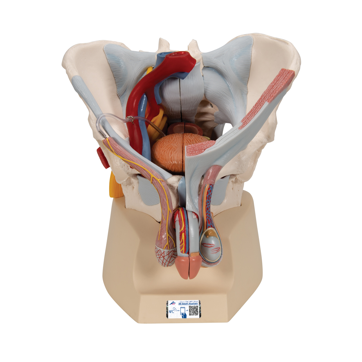
Male Pelvis Skeleton Model With Ligaments Vessels Nerves Pelvic Floor Muscles Organs 7 Part 3b Smart Anatomy 1013282 3b Scientific H21 3 Genital And Pelvis Models Anatomical Models from www.3bscientific.com Arcus tendineus levator ani and the ischial spine Some of the most important include the major digestive organs, the intestines. The pubococcygeus (pc) muscle is the muscle that runs the show in pelvic floor health. 12 photos of the muscle anatomy pelvis. The levator ani is a broad sheet of muscle. There is very little movement of the pelvic girdle because of its connection with the sacrum at the base of the axial. Cross the ls joint onto the trunk 2. Schau dir angebote von anatomie auf ebay an.
The pubococcygeus (pc) muscle is the muscle that runs the show in pelvic floor health.
It's supplied by ventral rami of first and 2nd sacral nerves (s1, s2). It is composed of three separate paired muscles; The piriformis is a triangular muscle 1 on either side on the very front of the posterior wall of true pelvis. These muscles have attachments to the pelvis as follows: The structure of the pelvis supports the contents of the abdomen while also helping to transfer the weight from the spine to the lower limbs. The function of the pelvic floor is to help assist with child birth, prevent incontinence and support organs within the pelvis. The appendicular muscles of the lower body position and stabilize the pelvic girdle, which serves as a foundation for the lower limbs. The intestines are supported by a series of muscles known as the pelvic. They have several functions, including helping to support the pelvic organs. These muscles move the thigh toward the body's midline. The pelvic region holds major organs under its layers of muscles. The muscles of the pelvis, hip and buttock anatomical chart shows how each muscle in this area of the body works with the others, and the various minor systems within the major ones. Arcus tendineus levator ani and the ischial spine
It takes origin from the inner aspect of pelvis along a line extending from the body of the pubis to the ischial spine. Cross the hip joint onto the thigh/leg 3. The floor of the pelvis is made up of the muscles of the pelvis, which support its. 12 photos of the muscle anatomy pelvis. Continence, then pelvic muscle exercises may be effective.
Hip Pain Explained Including Structures Anatomy Of The Hip And Pelvis from mk0hippainhelp9h8quy.kinstacdn.com Puborectalis, pubococcygeus, and the iliococcygeus. These bones also act as attachments for many muscles and ligaments within the pelvis and lower limbs. Cross the ls joint onto the trunk 2. The levator ani is a broad sheet of muscle. The levator ani muscles consist of three. These muscles, including the gluteus maximus and the hamstrings, extend the thigh at the hip in support of the body's weight and propulsion. It is a broad flat muscle. The main focus of this article will be the pelvic floor muscles.
The piriformis is a triangular muscle 1 on either side on the very front of the posterior wall of true pelvis.
On the other hand, if portions of those muscles are irretrievably lost, for example, due to complete. Pelvic floor muscles located wholly within the pelvis Attached to the pelvis are muscles of the buttocks, the lower back, and the thighs. A proper kegel exercise means a full contraction and relaxation of the pc muscle. Continence, then pelvic muscle exercises may be effective. The adductor muscle group, also known as the groin muscles, is a group located on the medial side of the thigh. This mri male pelvis axial cross sectional anatomy tool is absolutely free to use. The pelvic girdle, also known as the hip bone, is composed of three fused bones: Psoas consists of a pair of deep muscles (psoas major and iliacus) located on each side of the pelvis in the abdomen. The pubococcygeus (pc) muscle is the muscle that runs the show in pelvic floor health. It consists of three parts; It can be described as one of the bodies diaphragms. These muscles, including the gluteus maximus and the hamstrings, extend the thigh at the hip in support of the body's weight and propulsion.
Included in this group are the adductor longus, adductor brevis, adductor magnus, pectineus, and gracilis muscles. Puborectalis, pubococcygeus, and the iliococcygeus. It can be divided into the greater pelvis and the lesser pelvis. They have several functions, including helping to support the pelvic organs. The pelvic diaphragm is a wide but thin muscular layer of tissue that forms the inferior border of the abdominopelvic cavity.
Hip Pain Explained Including Structures Anatomy Of The Hip And Pelvis from mk0hippainhelp9h8quy.kinstacdn.com It is a broad flat muscle. The hip bone, sacrum and coccyx. Cross the ls joint onto the trunk 2. The pelvic floor muscles provide foundational support for the intestines and bladder. The pelvis's frame is made up of the bones of the pelvis, which connect the axial skeleton to the femurs, and therefore acts in weight bearing of the upper body. The function of the pelvic floor is to help assist with child birth, prevent incontinence and support organs within the pelvis. The pelvic floor muscles include; Muscles of the pelvis the muscles of the pelvis and hip control the vast range of movement of the legs and torso.
It can be described as one of the bodies diaphragms.
It helps maintain erect posture, abducts the thigh, and rotates the thigh outward. Reviews the functional anatomy of the pelvic floor structures, the effects of age on urethral support and the urethral sphincter, and attempts to clarify The pelvic region holds major organs under its layers of muscles. Cross the hip joint onto the thigh/leg 3. It takes origin from the inner aspect of pelvis along a line extending from the body of the pubis to the ischial spine. The pelvic diaphragm is a wide but thin muscular layer of tissue that forms the inferior border of the abdominopelvic cavity. The pelvic girdle, also known as the hip bone, is composed of three fused bones: An important group of muscles in the pelvis is the pelvic floor. It separates pelvis from the perineum. On the posterior side they are the glutei and on the anterior side the hip muscles extending into the thighs. The intestines are supported by a series of muscles known as the pelvic. The pelvis consists of the sacrum, the coccyx, the ischium, the ilium, and the pubis. Puborectalis, pubococcygeus, and the iliococcygeus.


0 Komentar